京械注准20162700767

Imaging processing

workstation

Insight Vision Workstation(IVW)is an intelligent medical image auxiliary diagnosis system developed by SinoVision, which can provide comprehensive advanced processing and visual solutions to customers. The system improves diagnostic efficiency through powerful advanced visual tools and customized workflow, and assists doctors in completing high-precision medical diagnosis and decision-making.
Advanced 3D and 4D medical imaging post-processing.

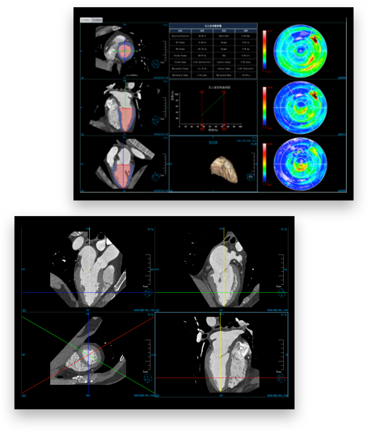
Cardiac coronary artery analysis

It can be used to analyze heart coronary artery related diseases and plaques, provide intelligent one-click diagnostic services and help doctors to quickly understand patients' pathological change.
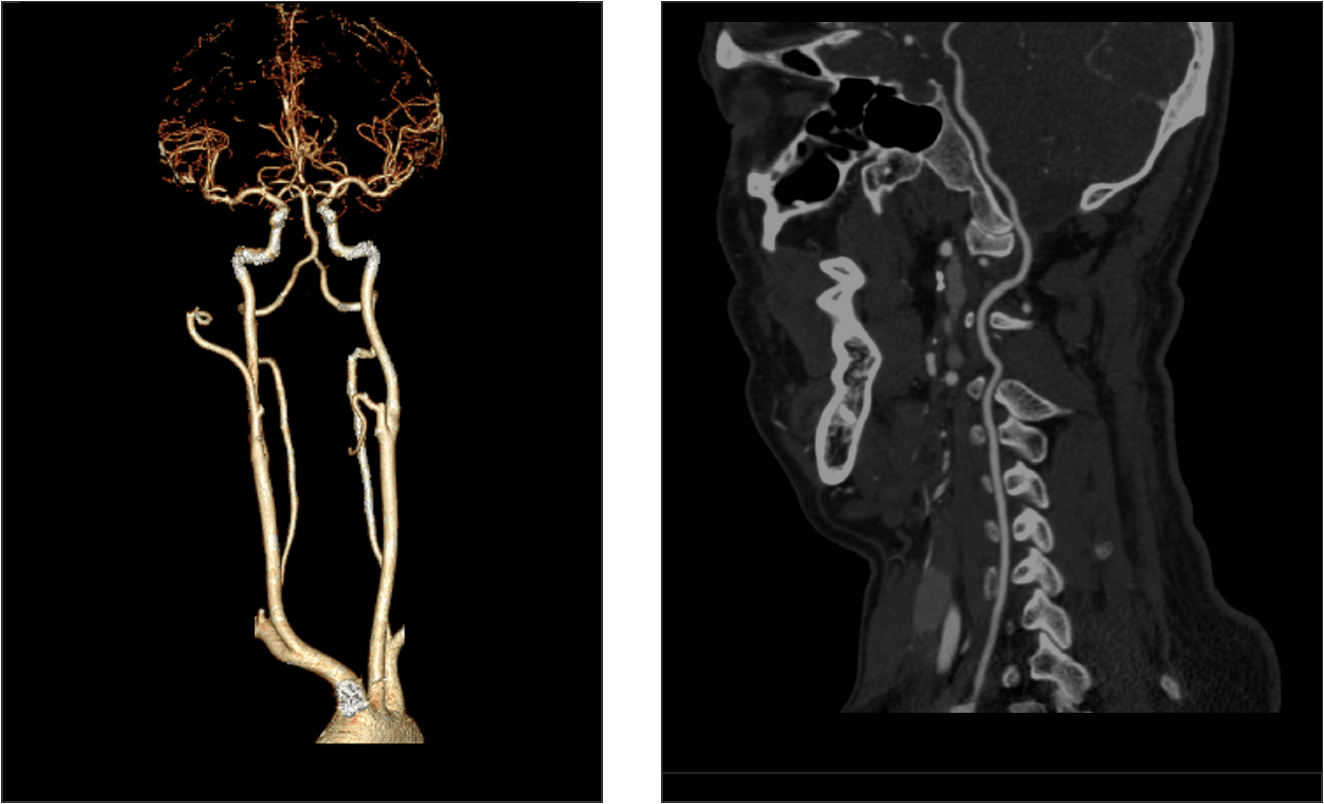
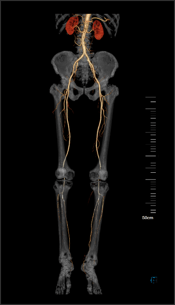
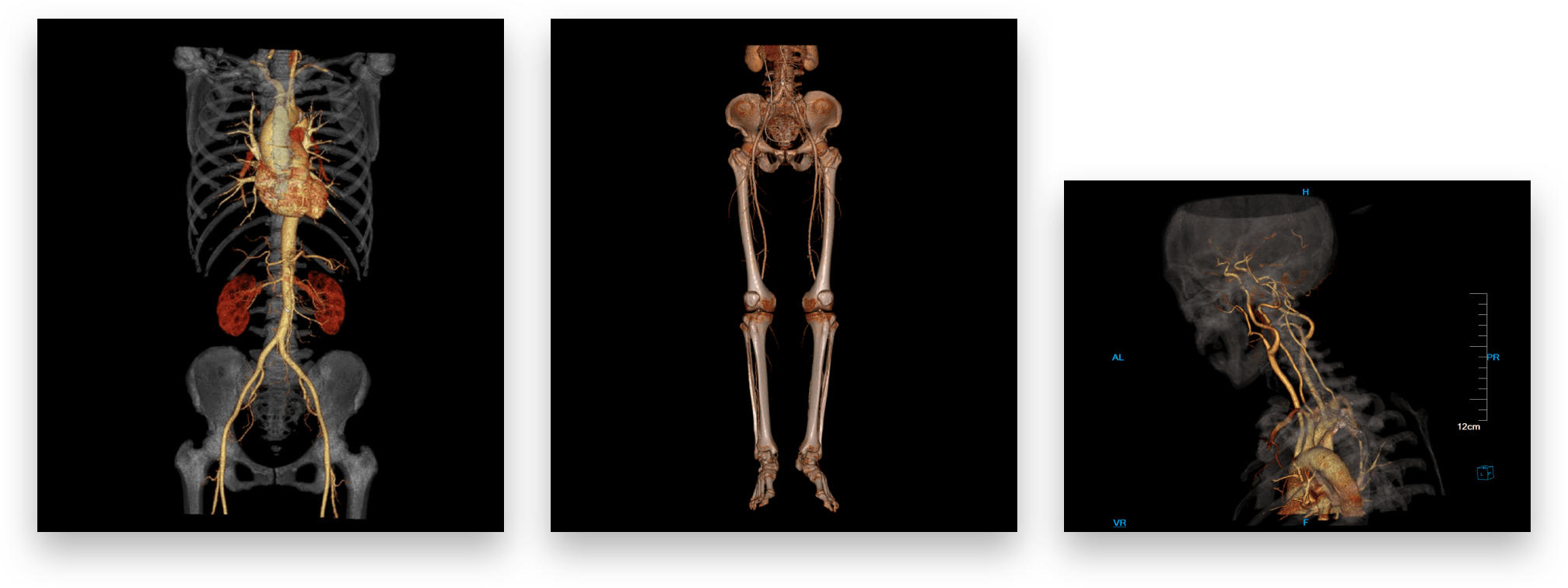
Cardiac calcium scoring

It supports the integral computation of calcified area of blood vessels, calculation of calcified volume, average CT value and maximum and minimum CT values of selected area and other functions.

Cardiac function analysis

To assist users in analyzing ventricular function and assessing whether there is hypofunction or dysfunction of the ventricle. To provide a variety of convenient interactive tools to assist doctors in more elaborate analysis and diagnosis.

FFRct

Coronary artery CT images based, the anatomy of coronary artery and the physiological assessment of coronary ischemia can be realized without the intervention of drugs and guidewire, and invasive examination is avoided.

CT digital subtraction angiography

Support subtraction of the data of head, neck, thorax and abdomen, extract blood vessels and analyze vascular straightening, stenosis and plaque.

Smart vascular intelligent analysis

Support full-automatic analysis of vascular diseases, including segmentation of vascular tissue, extraction of vascular centerline, automatic naming of vessels, stenosis detection and plaque component analysis, and provide diagnostic basis for atherosclerosis, aortic aneurysm and dissection.

Lung nodule analysis and follow-up

Support lung tissue extraction, automatically calculate three-dimensional volume, effective diameter and three-dimensional shape of pulmonary nodules, and provide the volume needed for nodule contrast. Follow-up visit of pulmonary nodules: support follow-up function of multi-phase imaging nodules, automatically calculate the volume change rate and doubling time of multi-phase pulmonary nodules after follow-up visit.

Lung function analysis

The lung function analysis software can extract pulmonary parenchyma and lung trachea, segment pulmonary lobe, extract emphysema and calculate the volume of emphysema in the lung. It is a powerful tool for clinicians to evaluate the structure and function of lung tissue and can quantitatively evaluate the change extent of the structure and trachea tissue of the lung with lesion by image.

Tumor analysis

Support the analysis of various tumors in human body, provide a variety of tools for tumor extraction, calculate relevant parameters of tumors according to WHO and RECIST standards and assist doctors in the analysis of tumors.

Brain perfusion analysis

To provide parameters such as cerebral blood flow volume, blood volume, mean transit time and time to peak, assist doctors in discovering cerebral ischemia tissue, make early diagnosis and prognosis of cerebral infarction and evaluate the effect of thrombolytic therapy.

Tumor perfusion

Tumor perfusion analysis software supports data correction, blood vessel definition, perfusion analysis and other functions. It can calculate parameters such as blood flow, blood volume, mean transit time, time to peak and surface permeability. These parameters can reflect micro vessel change of tumor angiogenesis in vivo, and help doctors to monitor the staging, grading and curative effect of tumors.

Hepatic perfusion

Hepatic perfusion analysis software supports data correction, blood vessel definition, perfusion analysis and other functions. It can calculate parameters such as hepatic arterial perfusion, portal venous perfusion, total hepatic perfusion, hepatic artery perfusion index, portal venous perfusion index and time to peak. These parameters can help doctors to identify and diagnose hepatocirrhosis, liver tumor and other lesions. It is of important value for qualitative diagnosis and differential diagnosis of hepatic diseases.

Virtual colonscopy

It can analyze the lesions on the inner wall of the colon, comprehensively observe the shape and size of lesions such as colon tumor and polypus, and assist doctors in locating and analyzing the lesions accurately.

Virtual endoscope

The application creates a three-dimensional model for displaying cavity and anatomical model graph in three-dimensional volume, and realizes flying-through function in the cavity. It support the modification of the depth of focus and multi-angle adjustment of viewing angle, and provide a variety of three-dimensional measurement tools.
  • Colonic endoscopic
  • Tracheal endoscopic
  • Endovascular sinogram

Dental analysis

Assist oral surgeons in making preoperative planning of false tooth implantation. Provide panoramic and cross-sectional images of teeth to help doctors to know the exact position and angle of implantation.

Multiple plane/ curve planar reconstruction

Support multi-dimensional and multi-angle plane and curve planar reconstruction of different tissues and organs, free adjustment of layer thickness and spacing, personalized customization and storage of reconstruction protocol, automatic browsing and storage of reconstruction batch processing, and various image processing tools.

3D visualization